Everything you need to know about age-related macular degeneration (AMD)

AMD and its different forms
A frequent pathology
Age-Related Macular Degeneration (AMD) is a chronic eye disease that most often appears after age 60 but can occur as early as age 50. The older you get, the higher the risk. Thus, it affects 1 in 2 people from the age of 80.
It is a deterioration of the central area of the retina, called the macula . AMD can take two main forms, each with different characteristics.
Atrophic or “dry” form
This is the least serious and most common form. It is characterized by a thinning of the macula which causes a slow and progressive decline in vision over a period of 5 to 10 years. Vision is blurred but color perception remains intact. This form can evolve into the second described below.
Exudative or “wet” form
This form is characterized by the development of abnormal blood vessels in the macula, called neovessels . The loss of vision is much faster than in the dry form and can range from a few days to a few months. In this form, the loss of vision can be more brutal (25% of cases).
What are the predisposing factors?
Beyond age , which represents the main risk factor, AMD can occur more readily in other contexts.Thus, the genetic predisposition is clearly identified . The risk of developing AMD is 4 times greater when a parent or sibling has it. Genes have been identified as implicated in the onset of the disease, such as the gene coding for complement factor H (a protein involved in immunity) or for HTRA1 (a protease). Indeed, variants of these genes are present in significantly higher proportions in people with AMD than in the general population (30% versus 10%).
Among the other factors highlighted with certainty, tobacco increases the risk in a ratio of 3 out of 6. Similarly, obesity multiplies the risk by 2.
On the other hand, oxidative stress is now a strongly suspected factor in the occurrence of AMD. A healthy lifestyle , especially food, would help prevent the onset of AMD.
Symptoms of AMD
The symptoms of AMD are polymorphic and variable in their intensity. Most often, one of the most characteristic signs is a distortion of images, objects and straight lines . The corresponding medical term is metamorphopsia .
Also, a black spot in the central part of the visual field may appear: the scotoma. All of this can be associated with a decrease in vision. The appearance of just one of these signs should lead to prompt consultation with an ophthalmologist.At the beginning of the disease, it can also be a difficulty in perceiving details. Finally, it may be a change in color vision.
Generally, AMD does not lead to complete loss of vision (blindness). Nevertheless, the various symptoms can considerably penalize the daily life, and this in a growing way with the evolution of the disease.
AMD diagnostic tools
The AMSLER Grid
Screening is a very important principle because treatment must be as early as possible.
The AMSLER grid is an easy-to-use tool designed to detect the first signs of AMD but also to clinically monitor the evolution of the pathology. The Amsler grid is made up of fine white grids on a black background which make it possible to highlight the anomalies and deficits of the central visual field.
For the evaluation with this tool, you have to keep your glasses on, stand at a distance of 35-40 cm from the grid, hide one eye then the other. If the person sees distorted lines, a decrease in contrast or an alteration of central vision, we must immediately suspect AMD.
Other complementary examinations
The diagnosis is made by an ophthalmologist after carrying out several examinations.
These examinations, painless, are generally 3 in number:
- Angiography : this defines the type of AMD by visualizing the vessels of the retina after injection of a contrast product;
- Optical coherence tomography called " OCT ": it allows to observe the structure of the eye;
- Retinography or photography of the fundus: it makes it possible to specify the lesions at the level of the macula.
Current treatments and therapeutic avenues
AMD is the subject of significant research on the one hand, to prevent the disease, and on the other hand to slow it down as effectively as possible. Early medical treatment is essential.
Treatment of the atrophic form of AMD
To date, there is no treatment that can stop the process that leads to the loss of thickness of the macula in this atrophic form because the damage to the retina is irreversible.
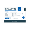
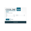
Nevertheless, the prescription of micronutrients in the form of food supplements has a braking interest on the evolution of the disease.
And moreover, as there is a risk of evolution towards an exudative form, it is necessary to set up a rigorous monitoring.
Treatment of the exudative form of AMD
The arrival of anti-VEGFs
Since 2006, the management of exudative AMD has experienced great progress thanks to the appearance on the market of treatments called anti-VEGF . This drug is directly injected into the eye after instillation of anesthetic eye drops.
Without curing the disease, repeated intravitreal injections of anti-VEGF prevent the formation of new vessels and reduce damage to the retina by drying out the associated edema. They have become the reference treatment and have thus made it possible to halve the rate of blindness linked to complications of AMD worldwide.
More efficient protocols
Since this discovery, the protocols have greatly evolved. Initially, these treatments were validated with systematic injection schemes. For example, an average of 7 injections per year has long been a benchmark. From now on, their frequency adapts more to each patient, to the rhythm of the progression of the neovessels.
In addition, promising research is emerging from several teams concerning gene therapy or injections of multipotent stem cells. These new therapies open the way to considering, in the future, treatments that are probably less restrictive than repeated injections of anti-VEGF (1) .
A healthy lifestyle and food supplements beneficial for vision health
Role of food for the retina and vision
For a long time, diet has been thought to have an influence on the disease. Indeed, there are beneficial nutrients in the diet whose consumption would have a protective effect . This is the case of omega 3, vitamins with an antioxidant function (vitamins C, E) as well as trace elements such as zinc for example.
Thus, a healthy and diversified diet can preserve and nourish the needs of the retina whose aging impoverishes the capacity of its tissues to maintain certain effective functions. According to a team from INSERM in Bordeaux, the adoption of a Mediterranean diet would contain adequate resources of nutrients necessary for retinal tissue (2) .
Dietary supplements offer proven efficacy
Proven study results
Given the impact of AMD on quality of life, researchers are studying all the actions to be taken to prevent or delay the development of the disease. Thus, the nutritional track has been studied and has shown that food supplements have proven efficacy in protecting and slowing the progression of retinal lesions.
The recommendations come from several large-scale studies, including the AREDS 2 study published in 2014 by an American team. The composition of food supplements has been determined through his studies (3) .
The ophthalmologist prescribes the intake of food supplements allowing supplementation with antioxidants and specific pigments.
Xanthophyll pigments: lutein and zeaxanthin
These two substances are part of the carotenoid family and have antioxidant properties. Indeed, lutein and zeaxanthin are major components of the macula.
The results of the AREDS 2 study show an 18% reduction in the progression of advanced AMD in subjects who received 10 mg of lutein and 2 mg of zeaxanthin (4) .
Omega-3s and especially DHA
The retina is schematically composed of the association of 2 elements: the neural retina and the pigmentary epithelium . The latter represents a protective coating that allows the passage of nutrients from the outside to the retina. It is particularly rich in polyunsaturated fatty acids (PUFA) and in particular in DHA (docosahexaenoic acid) which corresponds to a type of omega-3 (5) .
A study showed the effectiveness of a daily consumption of DHA on visual performance and in its preventive role on AMD (6) . The dose recommended by the European Food Safety Authority ( EFSA ), to take advantage of their benefits on vision, is 250 mg of DHA per day .
Vitamins and trace elements
The micronutrients involved in the protection against oxidative stress are well identified. Vitamins A, C and E are fundamental in this circuit, just like zinc, copper and selenium protect cells against oxidative stress. Taking antioxidant vitamins and trace elements in supplementation completes the identified needs (3) .
Other beneficial measures
Smoking cessation , maintaining gentle and regular physical activity and controlling a correct body mass index are all useful and complementary recommendations to prevent the onset and/or worsening of AMD.
Adaptations to improve the comfort of life for people suffering from AMD
Functional rehabilitation is one of the valuable complementary tools to help patients with AMD maintain their autonomy and continue to carry out activities of daily living in the best possible conditions.
Several professionals can subscribe to this objective. Among them, the orthoptist plays an essential role in this support: he will help to tame this loss of vision and find compensation mechanisms. It is indeed possible to learn how to make better use of one's residual visual abilities, relying in particular on peripheral vision. Also, the optician specializing in low vision will help advise the most appropriate visual aids. The occupational therapist can intervene in several areas such as the implementation of arrangements and adaptations of the place of life. The list of speakers is not exhaustive and other professionals may also be called upon (psychologist, psychomotrician, etc.).
In the end, AMD requires multidisciplinary care. Significant progress has been made in recent decades through research. Not only are ophthalmological treatments in full development, but the field of food and food supplements are proving essential to prevent and slow the progression of the disease.
Sources:
1. Age-related macular degeneration (AMD) ⋅ Inserm, La science pour la santé [Internet]. Inserm. Link here
2. Reduce the risk of AMD by adopting a Mediterranean diet. Inserm, Science for health [Internet]. Link here
3. Agron E, Mares J, Clemons TE, Swaroop A, Chew EY, Keenan TDL. Dietary nutrient intake and progression to late age-related macular degeneration in the Age-Related Eye Disease Studies 1 and 2. Ophthalmology. 2021 Mar;128(3):425-42.
4. Chew EY, Clemons TE, Agrón E, Sperduto RD, SanGiovanni JP, Davis MD, et al. Ten-Year Follow-up of Age-Related Macular Degeneration in the Age-Related Eye Disease Study: AREDS Report No. 36. JAMA Ophthalmol. 2014 Mar 1;132(3):272‑7.
5. Querques G, Forte R, Souied EH. Retina and omega-3. J Nutr Metab. 2011;2011:748361.
6. Chong EWT, Kreis AJ, Wong TY, Simpson JA, Guymer RH. Dietary omega-3 fatty acid and fish intake in the primary prevention of age-related macular degeneration: a systematic review and meta-analysis. Arch Ophthalmol Chic Ill 1960. 2008 Jun;126(6):826‑33.
Share
